Picture Of Forearm Muscles And Tendons - Elbow Anatomy - MKS : Posterior compartment muscles of the forearm.. While the ventral side of the forearm is not exactly less complicated than the dorsal side, it appears. Vintage forearm muscle and tendon anatomical hand drawing. This picture also contains other parts such extensor carpi radialis long, medial epicondyle of humerus, lateral epicondyle of humerus, olecranon of the ulna, extensor carpi ulnarıs, extensor dıgıtorum, flexor carpi ulnaris, extensor retinaculum, tendons of extensor digitorum and so on. From superior to inferior, origin. This picture shows how they are inside your arm soft tissue generally means tendons and ligaments, although it is quite unusual to damage a ligament in.
A version of this midel is available to buy in the sketchfab store: In the anterior compartment, they are split into three categories: This type of forearm grade iii strain of forearm muscle: The posterior compartment of the forearm (or extensor compartment) contains twelve muscles which are chiefly responsible for extension of the wrist and digits, and supination of the forearm. Do it yourself as shown in the picture!
Deep muscles of the forearm and elbow - Netter | Upper ... from i.pinimg.com It originates from the lateral epicondyle of humerus via the common extensor tendon. Also, pollicis means thumb in latin. A square shaped muscle found deep to the tendons of the fdp and fpl. 12 (4 superficial + 3 mobile wad + 5 deep). Originates from the anterior surface of the ulna and attaches to the. This type of forearm grade iii strain of forearm muscle: Anconeus muscle is a small muscle that is triangular in shape. Tusindvis af nye billeder af høj kvalitet tilføjes hver dag.
A version of this midel is available to buy in the sketchfab store:
Most commonly it is the tendon of the extensor carpi radialis brevis muscle that is weakened or torn from injury or overuse. Tendons are fibrous cords, similar to a rope, and are made of collagen. There are many muscles in the forearm. Lesson on the anatomy of the forearm: Hold your elbow with thumbs up and other 4 extension of index finger. This type of forearm grade iii strain of forearm muscle: Originates from the anterior surface of the ulna and attaches to the. They control movements of the wrist, hand, fingers and thumb. A version of this midel is available to buy in the sketchfab store: The forearm muscle strains are graded into three categories which are described below: Cross sectional anatomy of the upper limb : One of these originated from the extensor carpi radialis brevis, became tendinous and travelled between the two radial extensor tendons. They receive additional fibers from the deep fascia of the forearm near the elbow, and from the septa which pass from this fascia between the individual muscles.
Hold your elbow with thumbs up and other 4 extension of index finger. Antagonist of forearm flexors ( bra… flexion powerful of elbow and supination of forearm; Most of these originate from the lateral epicondyle. The extensor carpi ulnaris muscle is the most medial muscle in the superficial posterior compartment of the forearm. Tendons are fibrous cords, similar to a rope, and are made of collagen.
掌長肌(Palmaris Longus muscle, PL) - 小小整理網站 Smallcollation from lh3.googleusercontent.com They receive additional fibers from the deep fascia of the forearm near the elbow, and from the septa which pass from this fascia between the individual muscles. Lesson on the anatomy of the forearm: Most of these originate from the lateral epicondyle. A tendon is the fibrous tissue that attaches muscle to bone in the human body. From superior to inferior, origin. Tendons are attached to muscles and to bone. Cross sectional anatomy of the upper limb : Originates from the anterior surface of the ulna and attaches to the.
In the anterior compartment, they are split into three categories:
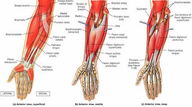
They control movements of the wrist, hand, fingers and thumb. They receive additional fibers from the deep fascia of the forearm near the elbow, and from the septa which pass from this fascia between the individual muscles. Antagonist of forearm flexors ( bra… flexion powerful of elbow and supination of forearm; The forearm is the region of the upper limb between the elbow and the wrist. The muscle fibers then descend towards the wrist area where they converge onto a narrow tendon. Cross sectional anatomy of the upper limb : The extrinsic hand muscles originate in the forearm and insert on structures within the hand. Tendons are under extreme stress when muscles pull on them, so they are very strong and are woven into the coverings of both muscles and bones. One of these originated from the extensor carpi radialis brevis, became tendinous and travelled between the two radial extensor tendons. Posterior compartment muscles of the forearm. These types of strains are quite severe and involve complete rupture of the muscle fibers and tendons. It originates from the lateral epicondyle of humerus via the common extensor tendon. Hold your elbow with thumbs up and other 4 extension of index finger.
Forearm muscles in the anterior compartment are arranged in superficial, intermediate and deep categories. Vintage forearm muscle and tendon anatomical hand drawing. While the ventral side of the forearm is not exactly less complicated than the dorsal side, it appears. One of these originated from the extensor carpi radialis brevis, became tendinous and travelled between the two radial extensor tendons. Find stockbilleder af forearm muscles tendons i hd og millionvis af andre royaltyfri stockbilleder, illustrationer og vektorer i shutterstocks samling.
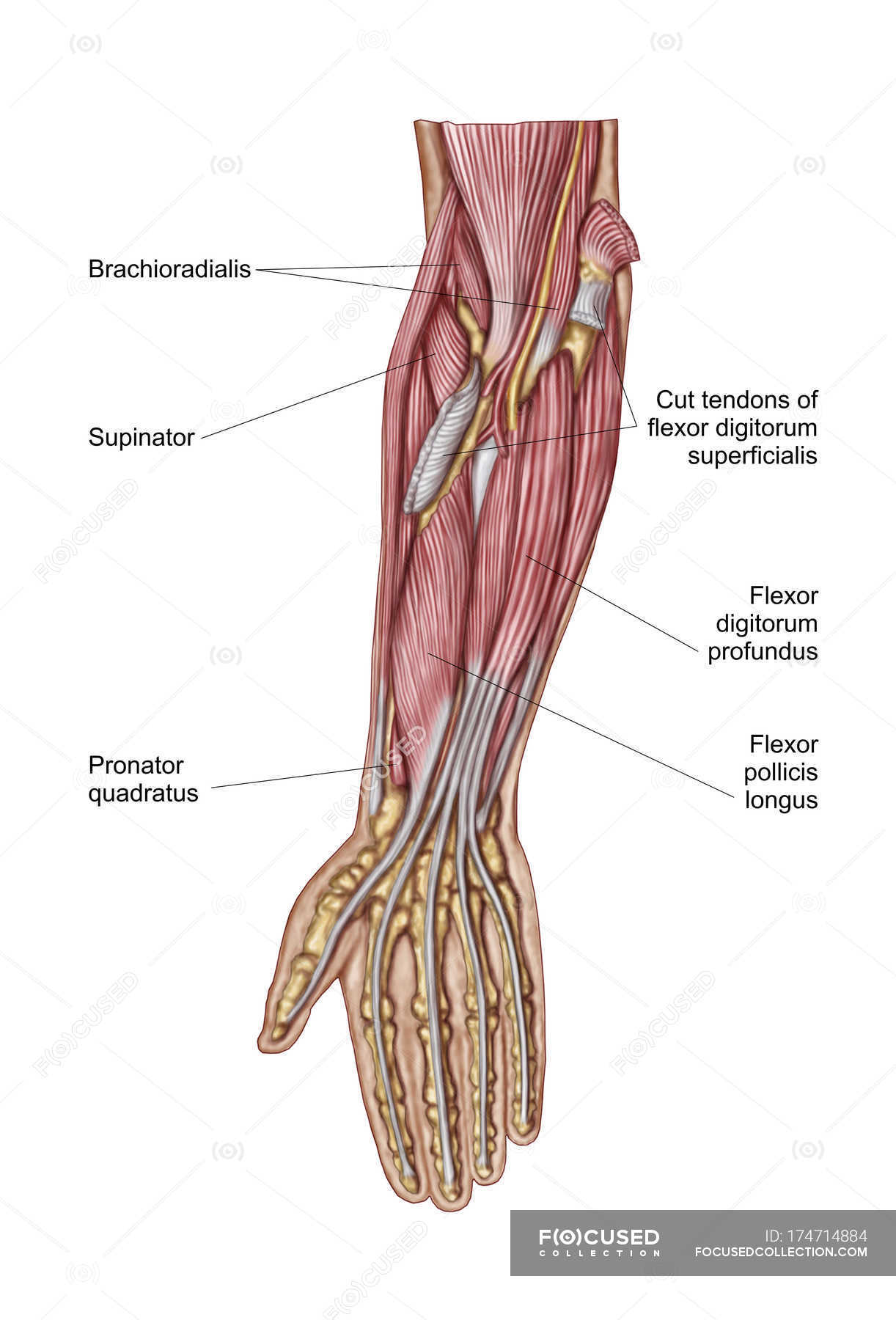
Anatomy of human forearm muscles with labels — Stock Photo ... from st.focusedcollection.com If you keep your hand flat on a table and. All superficial muscles are arises from the medial epicondyle of humerus but they are inserted into the different part except. There are many muscles in the forearm. Originates from the anterior surface of the ulna and attaches to the. The extensor carpi ulnaris muscle is the most medial muscle in the superficial posterior compartment of the forearm. Anconeus muscle is a small muscle that is triangular in shape. A version of this midel is available to buy in the sketchfab store: Tendons are under extreme stress when muscles pull on them, so they are very strong and are woven into the coverings of both muscles and bones.
Most commonly it is the tendon of the extensor carpi radialis brevis muscle that is weakened or torn from injury or overuse.
The muscles of this group take origin from the medial epicondyle of the humerus by a common tendon; When identifying the function of the forearm muscles, it is important to note that any forearm compartment muscle that crosses the elbow joint will act at this joint. The muscles of the upper arm are responsible for the flexion and extension of the forearm at the elbow joint. Posterior compartment muscles of the forearm. Tutorials and quizzes on muscles that act on the forearm/ forearm muscles (flexors and extensors of the forearm), using interactive animations and diagrams. Antagonist of forearm flexors ( bra… flexion powerful of elbow and supination of forearm; There are many muscles in the forearm. The pronator teres has two heads of. Also, pollicis means thumb in latin. A tendon is the fibrous tissue that attaches muscle to bone in the human body. Find stockbilleder af forearm muscles tendons i hd og millionvis af andre royaltyfri stockbilleder, illustrationer og vektorer i shutterstocks samling. It inserted independently into the. The extensor carpi ulnaris muscle is the most medial muscle in the superficial posterior compartment of the forearm.
Originates from the anterior surface of the ulna and attaches to the picture of forearm tendons. It originates from the lateral epicondyle of humerus via the common extensor tendon.


0 Komentar